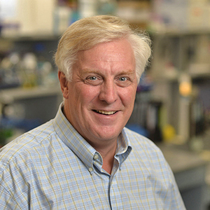
David R. Hyde Professor; Kenna Director of the Zebrafish Research Center

Research Interests:
My lab studies regeneration of retinal neurons from resident Müller glia, which are a class of radial glia that acts as adult stem cells in zebrafish. This is in contrast to the damaged human retina, where the resident Müller glia undergo gliosis to initially slow further retinal damage and often results in a glial scar. Thus, understanding how the zebrafish regenerates lost neurons may lead to therapies to treat several human neurological diseases, such as macular degeneration and retinitis pigmentosa in the retina or Alzheimer’s and Parkinson’s disease.
In response to a variety of different damage models, the loss of retinal neurons induces the resident Müller glia to dedifferentiate/reprogram and reenter the cell cycle to produce a neuronal progenitor cell (NPCs) through asymmetric cell division. These NPCs continue to proliferate and migrate to the damaged retinal layer and preferentially differentiate into the neuronal class that was lost. Because these Müller glia-derived NPCs can differentiate into any type of retinal neuron, they must be pluripotent. Because the regeneration preferentially differentiate into the neuronal cell types that were lost and they are restored to approximately the same number that were lost, signals must stimulate the initiation of the regeneration response, regulate the extent of Müller glia and NPC proliferation, and communicate the type and number of retinal neurons that were lost.
My lab is exploring a number of transcription factors and signaling pathways that are involved in the initiation of Müller glia reprogramming and proliferation and NPC proliferation. We identified that dying retinal neurons produce tumor necrosis factor-alpha (TNFα) to stimulate Müller glia proliferation, while Notch, which represses Müller glia proliferation, must be attenuated. In fact, injection of TNFα and the gamma secretase inhibitor R04929097 (to block Notch signaling) is sufficient to stimulate nearly 90% of the Müller glia to reenter the cell cycle to produce NPCs that amplify in number and differentiate into retinal neurons, all in the absence of retinal damage. Thus, expression of TNFα and repression of Notch signaling are necessary and sufficient to stimulate a regeneration response. We also determined that Sox2 is required and sufficient for Müller glia reprogramming. These three proteins regulate the expression and activation of two transcription factors that are both necessary for Müller glia proliferation, Ascl1a (Achete scute-like 1a) and Stat3. While Stat3 is expressed in all Müller glia in the light-damaged retina, Ascl1a is only expressed in proliferating Müller glia. It is unclear what regulates the differential expression patterns of Stat3 and Ascl1a during retinal regeneration. Finally, the Hippo-Yap and NF-kB pathways play critical roles to initiate the regeneration response. We are very interested in the relationship of these different signaling pathways to properly regulate retinal regeneration.
Recently, we initiated a new project to examine the transcriptomics and epigenomics of Müller glia during the initial stages of retinal regeneration. Using FACS to isolate Müller glia in response to different damage models, and a combination of RNAseq and single-cell RNAseq, we are examining what are the common and different gene expression patterns to identify the general molecular mechanisms and the specific mechanisms required to regenerate different classes of retinal neurons. Using ATACseq to identify changes in the compaction of the genome, we are studying the relationship of chromatin changes with gene expression changes to reveal the potential transcription factors required for Müller glia reprogramming and reentry into the cell cycle. In collaboration with collaborators at Johns Hopkins School of Medicine, Ohio State University and University of Florida, who are performing analogous experiments in the chick and mouse, we hope to identify the molecular changes (both chromatin and gene expression) that allow Müller glia reprogramming and regeneration in zebrafish, but not in the chick or mouse. As we identify potential key candidate molecules, we will use a variety of genetic and cell biological approaches to test the function of each candidate in both zebrafish and mouse, with the goal of identifying a the necessary cocktail of expression changes that are required for Müller glia-dependent retinal regeneration in mouse and ultimately humans.
We also initiated a project to study the effects of blunt force trauma on the zebrafish brain. Focusing on the damaged cerebellum, we determined that the blunt force trauma results in the death of neurons, which are subsequently regenerated. Manipulating the force of the trauma allowed us to develop a mild, moderate, and severe damage models. We are currently investigating the source of the regeneration. In addition, we determined that the trauma results in both a learning deficit and loss of short-term memory, while long-term memory is not significantly affected. We intend to apply our knowledge of retinal regeneration to study the mechanisms underlying neuronal regeneration in the damaged brain.
Biography:
- Director of the Center for Stem Cells and Regenerative Medicine 2014-Present
- Howard J. Kenna, C.S.C. Memorial Director of the Center for Zebrafish Research 2001-Present
- Professor, Department of Biological Sciences, University of Notre Dame, IN 2000-Present
- Director, University of Notre Dame Zebrafish Research Facility, IN 1995-2001
- Associate Professor, Department of Biological Sciences, University of Notre Dame, IN 1995-2000
- Assistant Professor, Department of Biological Sciences, University of Notre Dame, IN 1988-1995
- Senior Research Fellow, Division of Biology, California Institute of Technology, CA 1988
- Postdoctoral Research Fellow, Division of Biology, California Institute of Technology, CA 1985-1988
- D. Biochemistry, Pennsylvania State University, PA 1985
- S. Biochemistry, Michigan State University, MI 1980
Recent Papers:
- Campbell, L J, and D R Hyde. “Opportunities for CRISPR/Cas9 Gene Editing in Retinal Regeneration Research.” Advances in Pediatrics., U.S. National Library of Medicine, 23 Nov. 2017.
- Lahne, M, and D R Hyde. “Live-Cell Imaging: New Avenues to Investigate Retinal Regeneration.” Advances in Pediatrics., U.S. National Library of Medicine, Aug. 2017.
- Gorsuch RA, Lahne M, Yarka CE, Petravick ME, Li J, Hyde DR. “Sox2 Regulates Müller Glia Reprogramming and Proliferation in the Regenerating Zebrafish Retina via Lin28 and Ascl1a.” Advances in Pediatrics., U.S. National Library of Medicine, Aug. 2017.
- Lahne M, Gorsuch RA, Nelson CM, Hyde DR. “Culture of Adult Transgenic Zebrafish Retinal Explants for Live-Cell Imaging by Multiphoton Microscopy.” Advances in Pediatrics., U.S. National Library of Medicine, 24 Feb. 2017.
- Houbrechts AM, Vergauwen L, Bagci E, Van Houcke J, Heijlen M, Kulemeka B, Hyde DR, Knapen D, Darras VM. “Deiodinase Knockdown Affects Zebrafish Eye Development at the Level of Gene Expression, Morphology and Function.” Advances in Pediatrics., U.S. National Library of Medicine, 15 Mar. 2016.
- Lahne M, Li J, Marton RM, Hyde DR. “Actin-Cytoskeleton- and Rock-Mediated INM Are Required for Photoreceptor Regeneration in the Adult Zebrafish Retina.” Advances in Pediatrics., U.S. National Library of Medicine, 25 Nov. 2015.
- Lahne, M, and D R Hyde. “Interkinetic Nuclear Migration in the Regenerating Retina.” Advances in Pediatrics., U.S. National Library of Medicine.
- Conner C, Ackerman KM, Lahne M, Hobgood JS, Hyde DR. “Repressing Notch Signaling and Expressing TNFα Are Sufficient to Mimic Retinal Regeneration by Inducing Müller Glial Proliferation to Generate Committed Progenitor Cells.” Advances in Pediatrics., U.S. National Library of Medicine, 22 Oct. 2014.